GI tract anomaly detection from endoscopy images
This research endeavors to develop comprehensive techniques for detecting, localizing, and segmenting anomalies within gastrointestinal (GI) organs, extending beyond single-organ focus. The primary aim is to address critical needs in medical imaging, particularly in endoscopy, by advancing anomaly detection and segmentation methodologies. Validation on publicly available benchmark datasets forms an integral part of this pursuit to enhance medical imaging procedures and ultimately improve patient outcomes.
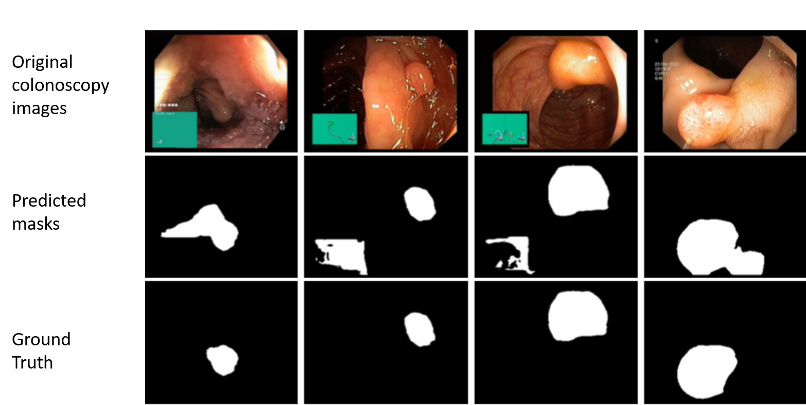
Problem:
- Detection of polyps in GI organs during endoscopic procedures is challenging.
- Variations in polyp shape, size, intensity, and specular reflections in colonoscopy images complicate their identification by endoscopists. This difficulty in detection not only impacts patient care but also contributes to higher mortality rates among colorectal cancer (CRC) patients.
- Need for accurate and efficient detection and segmentation techniques to address these challenges in endoscopic imaging.
Research Aim:
- Develop novel architectures for polyp segmentation from endoscopy images.
- Surpass the performance of existing state-of-the-art techniques.
- Provide advanced tools to support endoscopists in more effective anomaly detection during colonoscopy.
Outcome So Far:
- Initial investigations laid groundwork for exploring novel deep learning architectures.
- Preliminary results show promising advancements beyond existing techniques.
- Ongoing efforts focus on refining and optimizing methodologies for improved anomaly detection during colonoscopy procedures.
Teams: Bishesh Khanal, Sharib Ali, and Shruti Shrestha
Research Themes:
Computational Endoscopy, Surgery & Pathology (CESP), Transforming Global health with AI (TOGAI)
Project Category:
Computer Vision, Machine Learning, Medical Imaging